
X-ray Diagnosis
X-ray Diagnosis
Simple but sophisticated, welcome to the world of the digital revolution. The fastest and the most precise diagnosis thanks to the latest digital technology. With the exclusive 2d+ technology is much easier to record the focuses of the jaw with much more details rather than the simple record.
The use of the x-ray in dentistry is necessary, because it gives us a complete look in the health condition of the patients teeth.
The digital x-ray procedure differs from the analog x-ray procedure because of the newest and more sophisticated technology in radiological recording that gives an extraordinal quality of photo with minimum radiation. It gives a complete precision and safety for the patients.
EXTRAORAL X-ray DIAGNOSIS
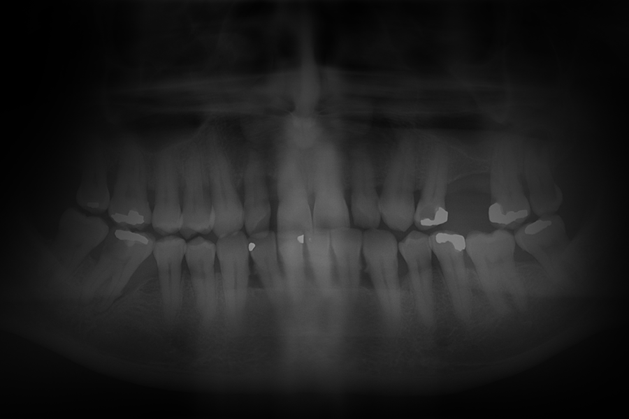
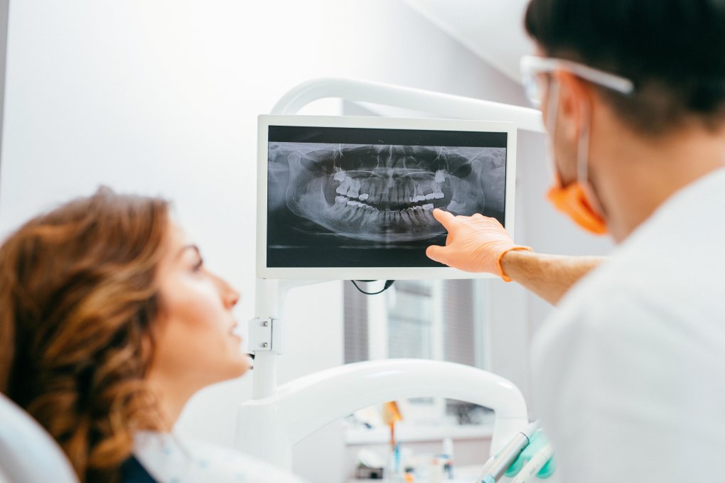
An orthopantomogram is an X-ray image of your whole mouth, including your upper and lower jaw. and teeth. The X-ray machine moves around your head while taking the image. This provides a. complete ear to ear image of your mouth and teeth.
During an OPG the patient remains in a stationary position (seated or standing) while both the x-ray source and film rotate in combination around the patient. The x-ray source rotates from one side of the jaw, around the front of the patient, and then to the other side of the jaw. The film rotates opposite to the x-ray source behind the patient. It takes a few seconds during which the patient must remain completely still.
This technique creates a panoramic image the should include the lower limits of the mandible, the upper limits of the maxillary sinuses, and the mandibular condyles and temporomandibular joints laterally. Panoramic image limitations include inherent anatomy distortion, double images, ghost images and do not provide a spatial relationship between structures.
INTRAORAL X-ray DIAGNOSIS
INTRAORAL X-ray DIAGNOSIS
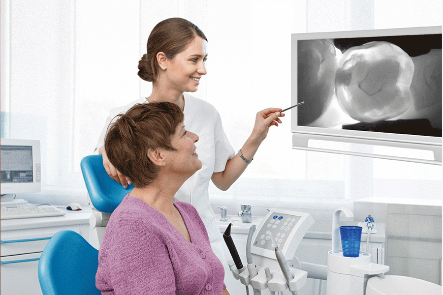
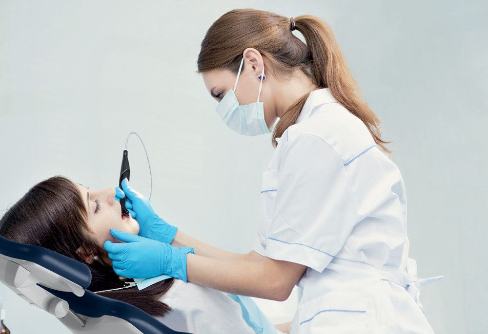
With the idea to give a new perspective on caries and to make the invisible visible, we introduce to you tomorrow’s technology today. It’s about the so-called laser detection of caries available only in our dental center “Dr.Batkoski”. It’s very important that this camera reveals those early stages of cavities before it has the opportunity to give any symptoms. The Kavo Diagno Cam diagnostic camera before is sure to reveal the caries at the earliest stage and enables you to correct your teeth with the principles of the modern, minimally invasive or as we say painless dentistry. Kavo Diagno cam uses healthy tooth structures as fluorescent light transmitters produced by the camera itself. At the same moment the laser releases the light, the video camera takes the moment of passing the beam through the tooth and transports it to the monitor, and the dentist and you can see it in real time and most importantly to understand the condition of your teeth. Such a device can detect caries where there is no apparent damage to the surface, and caries are hidden and can’t be felt or see by a regular probe with a probe.
What are the advantages of laser caries detection?
In addition to, of course, the discovery of saturated caries in their initial phase, the Diagnocam tool offers you the following benefits:
- Determines the diagnosis of the toothpicks
- Determines the caries under old seals.
- Monitor the caries and its development, data can be stored and situation monitored
- Offers minimal invasive treatment
- It saves a healthy tooth substance
- Opens the possibility of preventive action
- Does not emit harmful radiation as opposed to X ray machines
- Confident, without pain and increases the trust of the patient
- Treatment of early caries implies the selection of methods of minimally invasive dentistry where a tooth substance is saved, thus protecting healthy parts of the tooth and preventing further complications and treatment of the tooth, and the patient saves money for a longer time interval.
Contact us
General Inquiries
Address
-
Marko Nestoroski Street, No.147, Ohrid
Hours
-
Mon-Fri 9:00AM — 6:00PM